Actinic Keratosis vs. Melanoma Understanding the Difference
When a Sun Spot Raises Questions
A new spot on your shoulder. A mole that seems darker. A patch of roughness on your forehead that wasn’t there before. Our skin constantly reflects how we live—and how much time we spend under the sun. But not every growth or mark is harmless.
Two of the most commonly confused skin conditions are actinic keratosis and melanoma. Both involve damage from ultraviolet (UV) exposure, both can resemble one another in early stages, and both require careful cancer screening. Understanding how they differ is essential to protecting your skin health and preventing more serious skin cancer down the line.
The Core Difference: Cell of Origin and Early Skin Cancer Signs
At the biological level, actinic keratosis and melanoma arise from very different skin cells, leading to unique behaviors and outcomes.
Actinic Keratosis (AK) — Surface-Level Precancer
Keratosis actinic, commonly called AK, develops in keratinocytes, the main protective cells of the epidermis. Years of sunlight and ultraviolet radiation damage these cells’ DNA, causing abnormal growths that appear as dry, scaly patches. AKs are often referred to as precancerous lesions because they can evolve into squamous cell carcinoma, a type of skin cancer that develops more slowly but can invade deeper layers if ignored.
Melanoma — Deep, Aggressive Cell Mutation
Melanoma begins in melanocytes, the pigment-producing cells that determine skin tone. When UV light causes DNA mutations in these cells, they can grow uncontrollably and spread rapidly.
Although melanoma represents only about 1% of all skin cancers, it causes most skin cancer deaths due to its ability to metastasize.

https://www.everydayhealth.com/skin-cancer/what-are-the-different-types-of-skin-cancer/
(melanoma photo and carcinoma photo examples are often used by dermatologists during visual screenings to aid comparison.)
How Each Skin Cancer Condition Looks and Feels — Basal Cell and Squamous Cell Differences
When you compare actinic keratosis, melanoma, and other forms of skin cancer such as basal cell carcinoma, you start to notice key differences.
Below are common indicators to help you identify unusual moles, spots, or skin growths worth examining.
|
Feature |
Actinic Keratosis |
Melanoma |
|
Color |
Pink, red, or skin-colored |
Brown, black, blue, or multicolored |
|
Texture |
Rough, dry, or scaly patch |
Smooth or raised mole; may ooze or crust |
|
Borders |
Often indistinct |
Irregular, blurred, asymmetric |
|
Symptoms |
May itch, sting, or feel tender |
Sometimes itch or bleed; often asymptomatic |
|
Common Sites |
Face, scalp, ears, forearms, hands |
Anywhere, even non-sun areas; men = back, women = legs |
Basal cell carcinoma, though usually less aggressive, may appear as a pearly bump or waxy lesion that bleeds easily.
Squamous cell carcinoma, which may develop from actinic keratosis, typically looks like a thickened patch or raised sore that doesn’t heal.
(Always request professional skin imaging and cancer screening when you notice changes in moles or unusual skin textures.)
Shared Risk Factors: Sun Exposure, Skin Type, and
Cellular Damage
Both melanoma and actinic keratosis share several risk factors, but the degree and type of UV exposure often determine how skin cancer develops.
Major risk factors include:
-
Prolonged sun exposure without protection
-
Fair skin or light-colored eyes and hair
-
A personal or family history of skin cancer
-
Frequent tanning bed use
-
Weakened immune system
-
Outdoor professions such as construction or agriculture
-
Age over 50 (due to cumulative UV damage)
Basal cells, squamous cells, and melanocytes all respond differently to UV injury. In keratosis actinic, abnormal cells remain in the epidermis. In melanoma, the mutated cells invade deeper tissue layers. Over years, these patterns reveal why some individuals develop precancerous growths while others experience more aggressive malignancies.
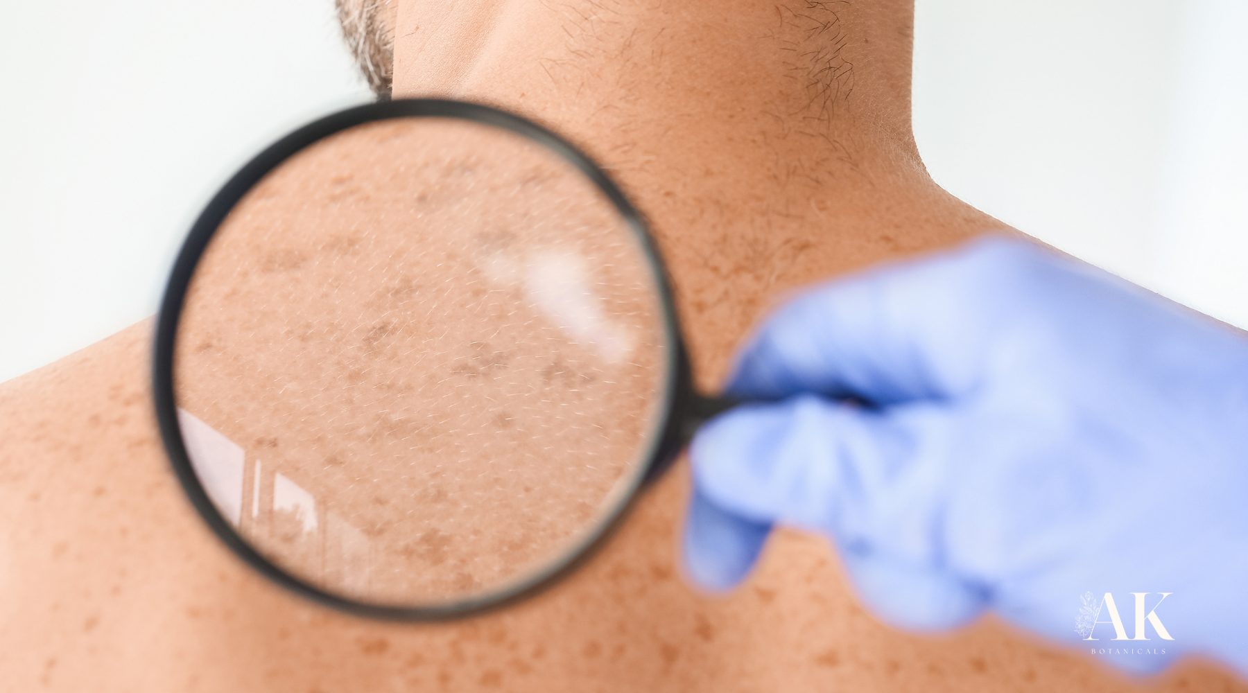
Cancer Screening and Skin Imaging: When to See a Dermatologist
If you notice unusual spots, moles, or persistent dry patches, don’t wait. Cancer screening and skin imaging technologies have made early detection far more accurate and less invasive.
For Actinic Keratosis:
Dermatologists may diagnose AK visually or use skin imaging tools like dermoscopy to assess surface texture. Some cases may require a biopsy for confirmation.
For Melanoma:
Dermatologists may photograph or digitally map moles over time using total-body skin imaging. Any irregular pattern, new growth, or rapidly changing color can trigger a biopsy for histological evaluation.
Regular checkups ensure that any suspicious skin growths are caught early—when treatment is most effective.
Treatment Options and Preventive Skin Care
Once diagnosed, treatment varies depending on if the condition is precancerous or malignant.
For Actinic Keratosis:
-
Cryotherapy: Freezing the lesion with liquid nitrogen.
-
Photodynamic Therapy: A light-sensitive compound targets abnormal cells.
-
Topical Creams: Certain prescribed creams help shed damaged skin.
-
Minor Excision: Removing thicker lesions that resist other treatments.
For Melanoma or Other Skin Cancers:
-
Surgical Removal: The most common treatment for confirmed melanomas.
-
Immunotherapy or Targeted Therapy: For advanced cases, these help the immune system recognize and fight abnormal cells.
-
Ongoing Cancer Screening: Regular follow-up visits to detect recurrence or new growths.
Supporting Healthy Skin Cells Naturally — A Holistic Path Forward
While dermatological treatment is essential for any diagnosed skin cancer, holistic care plays a complementary role in maintaining overall skin health.
Ingredients such as Annona muricata (soursop) and Sanguinaria canadensis (bloodroot)—studied for their antioxidant and cytotoxic properties—show promise in laboratory studies for supporting the skin’s natural renewal processes.
Disclaimer: These findings are based on laboratory studies. They do not imply that botanical compounds diagnose, treat, cure, or prevent disease. Always consult your dermatologist before starting any new topical or supplement regimen.
(For educational reference, dermatologists may use carcinoma and melanoma photo comparisons during consultations.)
AK Botanicals and AKti-Clear™: Natural Support for Sun-Exposed Skin
AKti-Clear™ from AK Botanicals is a botanically infused skincare solution formulated to support sun-exposed skin. It features natural plant compounds inspired by research on soursop and bloodroot, carefully developed to promote healthier skin.
Important Disclaimer: AKti-Clear™ is formulated to support overall skin wellness. It is not intended to diagnose, treat, cure, or prevent any disease, including actinic keratosis, melanoma, or other forms of skin cancer. For medical treatment, diagnosis, or advice, please consult a qualified dermatologist.
Early Awareness, Prevention, and Consistent Care
Actinic keratosis, melanoma, and other skin cancers remind us how delicate and powerful our skin truly is. Early cancer screening, UV protection, and attention to changing moles, spots, or skin growths can make a life-saving difference.
Healthy skin cells thrive on awareness, prevention, and thoughtful care—inside and out. Whether through professional dermatology visits or holistic botanical support, long-term wellness begins with daily attention to your skin’s story under the sun.


